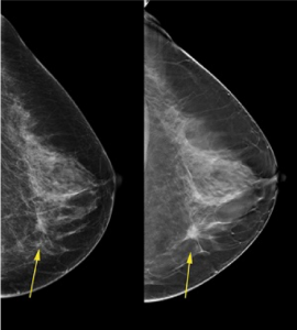
Conventional mammograms offer a two-dimensional (2D) view of breast tissue. The technology creates the 2D image from two X-ray images of each breast. Conventional mammograms are an effective tool to detect breast cancer and are currently the standard of care.
But three-dimensional (3D) mammography or digital tomosynthesis is proving to be even better—and the technology is now available at Welia Health.
Three-dimensional mammography combines 2D mammogram images with additional 3D views. Research studies show that 3D mammograms identify more cancers than traditional 2D mammograms. They also lower the number of false positives.
False positives occur when a mammogram shows an abnormal area that appears to be cancer, but ultimately is not. While false positives in the end are good (no cancer!), they lead to additional appointments, tests, and procedures—which means more mental, physical, and economic costs.
The benefits of 3D mammography
At Welia Health, we’re your partners in health. We equip ourselves with the best tools for your care, and a 3D mammogram is the latest advancement in breast imaging. Specifically, screening for breast cancer with a 3D mammogram can:

- Decrease need for follow-up imaging: A 3D mammogram machine creates both 3D and 2D images. Both types of images add value. Because the 3D image is an enhanced view of the breast tissue, radiologists can examine any abnormalities in closer detail without follow-up imaging.
- Better detect breast cancer in dense breast tissue: Breasts consist of milk glands, (glandular tissue), milk ducts, and fatty and fibrous connective tissue. The latter give breasts their size and shape and hold the other tissues in place. Breasts that have a lot of fibrous or glandular tissue, but not much fat, are considered dense. Both dense breast tissue and cancers appear white on a standard mammogram, which may make breast cancer more difficult to detect in dense breasts. But a 3D image of dense breast tissue enables physicians to see beyond the dense areas of tissue.
- Increase in cancer detection compared to 2D mammography: A 2013 prospective randomized study (or one that examines a group of people or cohort randomly) compared 3D and 2D mammography in more than 12,000 patients. Researchers determined a 27 percent increase in cancer detection with 3D mammography. Another 2013 study of more than 7,000 patients showed an increase in cancer detection of 50 percent.
Scheduling a mammogram
In general, Welia Health physicians recommend that our patients start having annual mammograms at age 40. However, medical history and individual breast cancer risk could make earlier screening prudent. If you’re concerned about breast cancer screenings or have questions about the procedure, you should reach out to your physician. Together you can determine what makes sense for you.
To schedule a mammogram, call Welia Health at 800.245.5671 or our Imaging Department at 320.225.3447. Three-dimensional mammograms are offered at Mora and Pine City facilities.


